


Sesamoid Surgery Should Be the Last Resort
This page provides a detailed explanation of seasamoid surgery. Surgery, however, should almost always be the very last resort for treating sesamoid injuries.
For a detailed guide to conservative treatment of sesamoid injuries read our Sesamoid Injury and Sesamoiditis Treatment Guide.
Sesamoid Anatomy
The two tiny kneecap-like sesamoid bones underneath the ball of your foot are housed inside a thick tendon called the flexor hallucis brevis. They sit inside the tendon similar to how a cherry pit sits inside a cherry, strongly attached to the surrounding tissue. In addition to the tendon, the sesamoids are held in place by many ligaments. These ligaments are strong and prevent the round sesamoid bones from rolling away from the ball of your foot when you put weight on them.
How Do You Injure Your Sesamoids?
Sesamoid injuries tend to occur when you place too much weight on the ball of your foot. The sesamoid bones are tiny round bones that are commonly injured during sports or even every day activities.
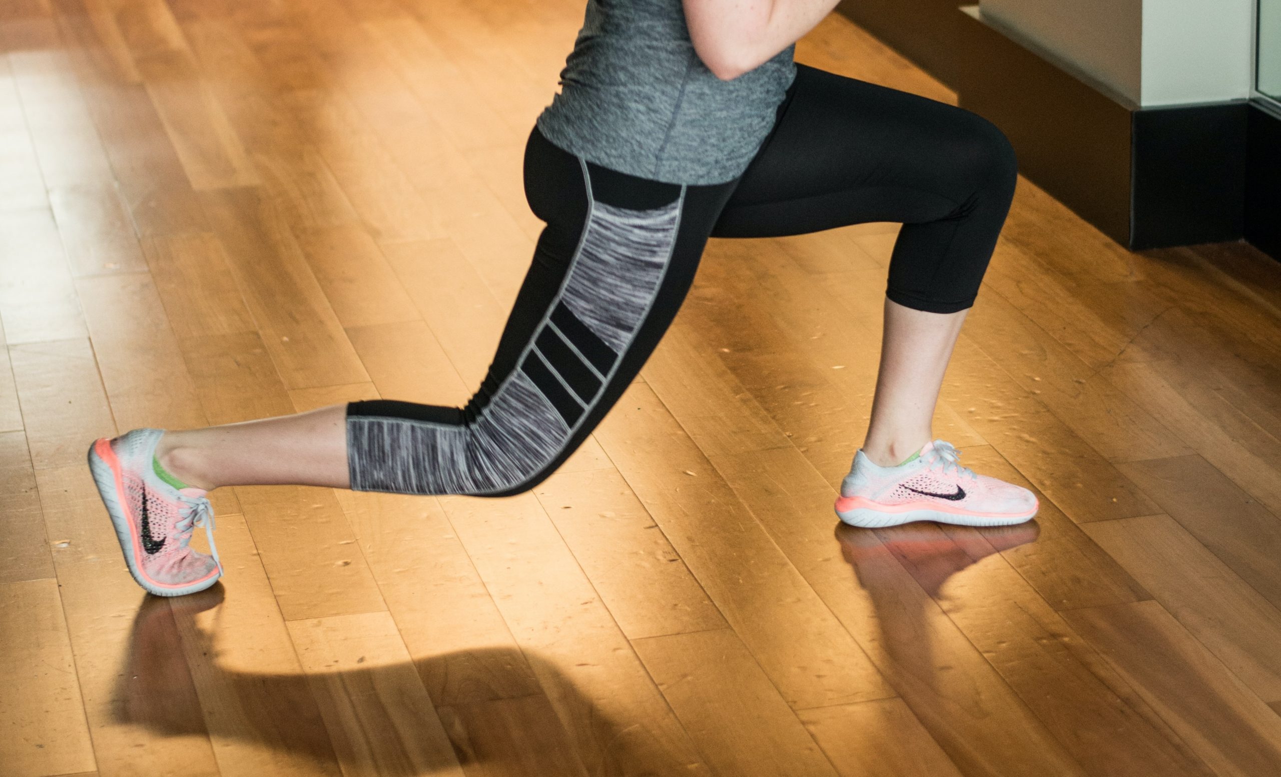 Common methods of injuring your sesamoids include:
- Lunging forward
- Landing after a jump
- Repetitive activities like walking or running
Your sesamoids can be irritated, which is called sesamoiditis. This generally responds to conservative treatment and rarely requires surgery.
Your sesamoids could also have a fracture, which causes pain. Sesamoiditis and sesamoid stress fractures occur in different ways, but can feel very similar. The sesamoiditis may be caused by more repetitive motion to the fall of the foot, while the fracture is caused by more high energy injury like running or jumping.
While most sesamoid fractures can be treated conservatively, they are more likely to need surgery than the sesamoiditis condition.
How do you tell the difference between a sesamoiditis and a sesamoid fracture?
Both the sesamoid fracture, and sesamoiditis:
- Can feel like a sharp or dull pain at the ball of the foot right under the joint
- Cause pain with movement of the big toe
- May be treatable by conservative methods, or require surgery
Mechanism of Injury:
- Sesamoid fracture usually has a sudden onset during an activity.
- Sesamoiditis usually has a gradual onset.
Imaging of the sesamoids:
Several techniques exist to evaluate the sesamoids. Below are some of the imaging techniques we use.
X-Ray:
The first technique is a regular x-ray. This may even be taken at your initial visit to the doctors office.
Fracture
- Fracture line (oblique line) in the sesamoid bone
- Both sesamoids are usually the same size but one has an oblique black line.
Sesamoiditis
- More common with bipartite sesamoid bone (meaning you were born with three sesamoids instead of 2 under your big toe joint)
- No fracture line noted.
You may have changes in the sesamoid bone, which can not be seen on the x-ray, yet will still cause pain. This is one of many reasons you may need additional imaging like a bone scan or an MRI.
Bone Scan:
A bone scan is a more advanced type of imaging that involves a very small amount of a radioactive substance, called a tracer, that localizes at the site of bone injury. This is generally performed for more difficult sesamoid injuries to determine exactly which sesamoid is injured.
We look for:
- Increased tracer localized around the sesamoid causing trouble
- Any additional areas of tracer uptake, indicating a bigger problem than sesamoid injury
If the bone scan identifies a specific area of tracer uptake in one sesamoid, it’s clear that that is the sesamoid causing trouble. Once the trouble sesamoid is located, it may be time to perform surgery to remove the sesamoid.
After the bone scan an MRI may be utilized to further evaluate the area.
MRI:
An MRI (magnetic resonance imaging) uses magnetic fields and the different tissue textures in your body to identify changes like swelling, infection, and damage to the bones and soft tissues. The MRI helps to distinguish injury of the joint capsule, tendon, and ligaments around the sesamoids from injury to the sesamoid bones themselves.
We look for:
- Areas of increased brightness in the joint, soft tissues, and ligaments
- Tears in the soft tissues
- Signals from inside the bone indicating edema (swelling) inside the bone
- Visible fractures of the sesamoid bones
An MRI can provide a definitive diagnosis and guide surgical planning.
When to Consider Surgery:
If you have already tried several months of conservative treatment including offloading in a surgical boot, special shoe modifications and custom orthotics with no relief, it may be time to consider surgery.
You should only consider having surgery if you are experiencing severe discomfort that doesn’t respond to any other treatments. Surgery is a big step, and it should always be your last resort for handling your sesamoid pain.
Be sure to see our Pre Surgical Preparation page for more details about planning your surgery and getting ready for your surgery.
The Sesamoid Removal Surgery:
If you have continued pain after months of conservative treatment for sesamoiditis or for a sesamoid fracture, the surgical option is to remove the troublesome sesamoid. Removing a sesamoid can cause several complications, which is why we try to avoid this surgery whenever possible.
During the surgery:
- Your surgery will start the same as any other foot surgery, detailed on our Pre Surgical Preparation page. You will be lying comfortably on your back for this surgery.
- A small 2-4cm incision will be made on the bottom, sometimes on the side, of your foot directly over the painful sesamoid. Important not toplace the scar on the outside the sesamoid bone since this may lead to painful scarring.
- Your surgeon will dissect down to the tendon which houses the sesamoid and remove part or all of the sesamoid from the tendon encasement.
- We use extreme caution when removing the sesamoid to preserve the tendon, which helps your foot function better and reduces complications after your surgery.
- An x-ray machine may be used during the surgery to help identify any pieces of the sesamoid that may have broken off during surgery.
- Once the sesamoid is removed, the skin is closed with stitches and a bandage is placed over the incision site.
Risks and Potential Complications of Sesamoid Surgery
There are certain risks that come with any foot surgery, these include the risk of bleeding, continued pain, infection, deep vein thrombosis, and the wound failing to heal. To read about each of these conditions and ways to identify them and avoid them we highly suggest you look at our Pre Surgical Preparation page and read about them in detail.
There are several risks that are specific to sesamoid removal surgery. A list of the most common risks are below:
- The biggest risk with sesamoid removal is continued pain after the surgery. Removing the sesamoid should stop the pain you feel, however this is not always the case. In some circumstances the pain continues after the sesamoid is removed, and additional conservative or possibly surgical procedures are necessary.
- A thick scar could develop on the bottom of your foot. The incision site is located on the bottom of the foot to allow the best possible view of the sesamoid. The incision may leave a thick scar that can be painful.
- We minimize the risk of thick scar development by using special dissection and stitching techniques for the bottom of the foot.
- You can minimize the risk of developing a thick painful scar by avoiding walking on the foot until your doctor approves weight bearing. Some physicians suggest vitamin E oil on the scar starting 3 weeks after your surgery. In severe cases an overnight silicone scar reduction pad can be used, if this is necessary your doctor will guide you to the correct place to purchase this.
- Your big toe can change directions. When you remove a sesamoid bone, part of the support system that keeps your big toe pointing straight forward is removed. Depending on which sesamoid bone is removed, your big toe might point away from the little toes (Hallux varus), or it might point toward the little toes (Hallux valgus, a bunion).
- We minimize the risk of big toe changes by carefully preserving the flexor hallucis brevis tendon which holds the sesamoid. In our experience we have seen very little deviation of the big toe when the tendon is properly protected.
- You can minimize the risk of your big toe changing directions by wearing custom made orthotics and supportive shoe gear after your surgery. It is also important not to touch the bandages after your operation.
I need a sesamoid surgery, what next?
At the Foot and Ankle Center of Washington we are experts at handling your sesamoid injuries through every step of the treatment. Please make an appointment with us at our clinic in Seattle so we can start building your treatment plan!
First you’ll establish a relationship with a foot and ankle specialist, and have a thorough clinical exam, as well as advanced imaging such as a bone scan or MRI.
Next, you’ll determine if surgery is right for you, what surgery must be done, and when is the right time to do it. Visit our Pre Surgical Preparation page to get a better idea of what surgery requires, and how to prepare for it. This helpful page will also guide you through deciding when to have your surgery.
If you are in the Seattle area, please make an appointment at our convenient Seattle Clinic. We can work with you to treat your sesamoid and get you back on your feet. Our talented foot and ankle specialists Dr.Huppin and Dr.Hale have many years of experience with conservative treatment, focusing on avoiding surgery and rehabilitating your sesamoid injury.
If conservative treatment fails, our podiatric surgeon Dr.Hale has many years of experience in the sports medicine foot and ankle surgery field and can help you navigate your sesamoid surgery.