
Extra Bones and Accessory Bones of the Foot
By Larry Huppin, DPM
There are 26 bones in the normal human foot but some people have extra bones. These extra bones (or accessory bones) usually do not cause any problems and you may not even know that you have one.
Sometimes, however, they do lead to pain in the foot or ankle. If think you might have an accessory bone in your foot and it is causing pain, contact us today to schedule an appointment in our Seattle clinic. Even when they cause a lot of pain we can usually treat them conservatively. Surgery is almost never necessary and it is always our last resort, but in rare occasions the extra bone must be removed to relieve pain.
What is an Accessory Bone?
When we are born, our bones are mostly cartilage. Over the first years of life this cartilage becomes bone. Most of the bones in the foot start to convert to bone in two areas and they eventually merge into one. Sometimes, however, they do not merge together and you are left with an extra bone. Accessory bones can occur in many bones in the foot, but they occur most commonly in three areas:
- Off of the navicular bone on the inside of the foot (accessory navicular, os navicularis or os tibiale externum)
- Off of the back of the ankle bone (os trigonum)
- Off of the cuboid bone (os peroneum)
How Are Extra Bones in the Foot Diagnosed?
Usually an examination of your foot and x-rays are all we need to diagnose an accessory bone. Sometimes additional imaging such as an MRI is necessary. Our goal is always to provide you with the correct diagnosis in the most cost effective method possible.
Treatment of Accessory Foot Bones
In most cases treatment of accessory bones is focused on removing pressure and tension from the painful bone in order to relieve pain. In the sections below I will discuss treatment for specific accessory bones. The key point I want everyone to understand is that in most cases surgery is not necessary to treat these extra bones in the feet.
Os Navicularis (Accessory Navicular)
Video: Accessory Navicular Treatment Options
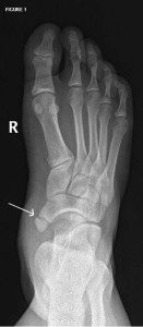
Accessory navicular
An accessory navicular is an extra bone or piece of cartilage located on the inside of the foot next to the navicular bone. It is inside the posterior tibial tendon which inserts into the foot in this area. This is sometimes also called os tibiale externum.
What Does an Accessory Navicular Look Like?
Many people with an accessory navicular will never know that they have one. But an os navicularis can present as a lump on the inside of the foot just off of the navicular bone. You can see on the x-ray that it just appears as an extra bone on the inside of the arch of the foot. Sometimes the skin over the accessory navicular becomes red and inflamed.
What Causes an Accessory Navicular to Hurt?
Sometimes accessory naviculars cause no pain at all and sometimes they can cause a lot of aching or sharp pain on the inside of the arch of the foot. There are several common problems that lead to pain:
- Trauma such as sprains of the ankle or foot.
- Pressure from shoes or boots: It’s common that stiff boots such as ski boots or hiking boots can irritate an accessory navicular.
- Flat feet: Many people who have accessory navicular bones also have flat feet. When the foot flattens (or pronates) too much, it puts tension on the posterior tibial tendon which can tug on the accessory navicular and lead to pain. In addition, as the foot pronates it can push the accessory bone into the side of the shoe.
- Overuse: Excessive walking or exercise can result in excessive pressure on these enlarged areas of the foot and cause pain.
Non-surgical Treatment of Accessory Navicular
Our primary goal when treating a painful accessory navicular is to reduce tension on the posterior tibialis tendon that inserts into the accessory navicular. We can almost always accomplish this with the use of properly prescribed foot orthotics.
If surgery has been recommended for treatment of this condition be sure to get a second opinion. We often see people with this condition with poorly made orthotics that are not adequate to relieve pain. In fact, if orthotics are made correctly, surgery is almost never necessary.
Be sure to see a podiatrist who specializes in orthotics before you have surgery. If you are in the Seattle area, contact us for an appointment.
Accessory Navicular and Flat Feet
If your foot is flat (which is common when an accessory navicular is present) we must make orthotics specifically designed for flat feet. Orthotics for treating an accessory navicular must:
- Reduce tension on the posterior tibialis tendon
- Reduce pressure on the accessory navicular
If your foot is flat then the orthotics must include special accommodations. Read about orthotics for flat feet here.
Home Treatment for Accessory Navicular
Below are our recommended home treatments for accessory navicular syndrome. You can try them for a few weeks but if you are still not better then see a foot and ankle specialist in your area. If your pain worsens see someone right away. The products below are the ones we recommend to our patients and they are also affiliate links so we may receive a small commission at no additional cost to you if your order from the link.
Use Arch Support in All of Your Shoes
In some situations a very supportive prefabricated arch support will reduce enough tension and pressure on the accessory navicular to provide 100% relief of pain.
Our preferred OTC orthtic for this problem is the FootChair Podiatrist Designed Orthotic. This  device works well for accessory navicular because of it’s relatively high arch, wide width and unique adjustable arch height.
For this condition we recommend using the highest arch that is comfortable for you in order to take tension off of the accessory navicular. The FootChair comes with pads that are added under the cover to increase the arch height so it will do a better job than most over the counter arch supports at reducing tension and pressure on the navicular.
For smaller shoes including high heels, flats and soccer cleats, the recommended OTC arch support is the FootChair Slim Orthotic with 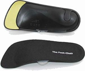 adjustable arch support. It’s the most effective OTC arch support we have found to fit in women’s heels and flats. It has the same exceptional adjustable arch support as the full-size FootChair but with a much slimmer profile. In addition it flexes to adapt to most heel heights.
When you are not wearing shoes use a sandal or flip-flop with very good arch support to reduce force on the accessory 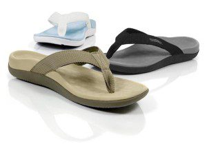
navicular. The sandals from Vionic are the best we have found for this situation.
Wear a Walking Boot to Allow the Tissue to Rest and Heal
If you are having a lot of pain you can try wearing a walking boot for about 10 days to start treatment. Wearing the 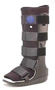 boot will eliminate almost all of the force on the posterior tibial tendon and on the navicular and let that tissue heal. After that go back into stable shoes with the arch supports to prevent the problem from returning.
You can see one of the better walking boot values here. We recommend wearing the arch support in the boot for best results.
Surgery for Accessory Navicular – the Kidner Procedure
In rare cases surgery might be necessary. The standard surgery for this condition is called a “Kidner procedure”.
This involves separating the tibialis posterior tendon from the navicular and removing the extra bone. The tendon is then reattached and the wound is sutured. Most patients are non-weight bearing (crutches or scooter) for a few weeks and back to most normal activities in about six weeks.
The use of orthotics after surgery is critical as properly made orthotics will reduce tension on the posterior tibialis tendon and pressure on the surgical site and allow for more rapid return to activity with less pain.
The Kidner procedure is usually very effective but we caution everyone who is considering this procedure that in most cases this condition can be treated without surgery. See us for a second opinion if surgery has been recommended.
If you are not in our area find a podiatrist near you who is extremely skilled in orthotic therapy as this skill will give you your best chance at avoiding surgery.
Treatment of Os Peroneum Pain
Video: Os Peroneum Pain Treatment Options
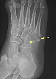
os peroneum
This accessory bone is found within one of the peroneal tendons off of the cuboid bone on the lateral (outside) portion of the foot.
Os peroneum are fairly common and are found in about 20% of the population. Those with an os peroneum are more prone to tendonitis and pain of the peroneus longus tendon on the outside of the foot. The goal of treatment is to reduce tension on the peroneal tendon as this almost always reduces pain.
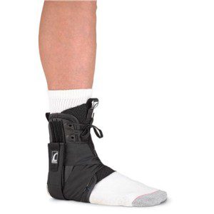
We use orthotics or arch supports to reduce tension on the peroneus longus tendon and in many cases this is all that is needed for treatment. In some cases we need to immobilize the tendon in a walking boot or ankle brace for a couple weeks first. Other treatments include icing and anti-inflammatory medications.
Orthotics for Os Peroneum
As with accessory navicular we use orthotics for treating chronic os peroneum pain, but it is a very different orthotic prescription than that used for treatment of the accessory navicular.
Orthotics for Os Peroneum should be prescribed to reduce tension on the peroneus longus tendon and to transfer pressure from the outside of the foot to the inside arch. In fact, this is a fairly difficult biomechanical problem and is a difficult problem to treat on your own.
We recommend you find a podiatrist who specializes in orthotics and biomechanics. If you are in the Seattle area contact us for an appointment. When properly made, orthotics are a very effective treatment for os peroneum pain.
Self Treatment of Os Peroneum Pain
Because this can be challenging to diagnose correctly, pain on the outside of the foot should be treated by a podiatrist or orthopedist to ensure proper treatment and avoid long term problems.
If you have pain on the outside of your foot that you think might be due to this problem, we recommend wearing a walking boot or ankle brace until you have a chance to get evaluated.
Treatment of Os Trigonum Syndrome
Video: Os Trigonum Syndrome Treatment
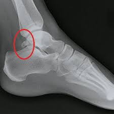
os trigonum
The os trigonum is an extra bone that occurs behind the ankle off of the back of the ankle bone (the talus). Most people don’t know they have an os trigonum since unlike the os navicularis and the os peroneum it cannot cause a lump on the outside of the foot.
Causes of Os Trigonum Syndrome
Os trigonum sometimes become painful – this is called os trigonum syndrome. In most cases os trigonum syndrome occurs following an injury such as an ankle sprain. This can also occur due to repeated downward pointing of the toes such as occurs during ballet.
A particular type of os trigonum injury is called the “nutcracker injury”. This refers both to the fact the the os trigonum is “crushed like a nut” between the ankle bone and the heel bone, and to the Nutcracker ballet.
Signs and Symptoms of Os Trigonum Syndrome
-
Deep, aching pain in the back of the ankle that occurs primarily when when pushing off of the big toe when walking or when pointing the toes downward
-
Tenderness to palpation of the area in the back of the ankle
-
Swelling in the back of the ankle
Diagnosis of Os Trigonum Syndrome
Os trigonum can mimic other conditions such as Achilles tendinitis and ankle sprains so it is important to get the correct diagnosis for the best treatment and results. In most cases a history, examination and x-rays will allow us to diagnose the condition. Sometimes additional imaging, such as MRI, is necessary.
Treatment of Os Trigonum: Non-surgical
In most cases surgery is not necessary and pain relief is achieved through treatments that can include a combination of the following:
- Rest and immobilization to allow the tissue to heal. Often we prescribe a walking boot to limit ankle motion but allow you to continue activity
- Ice to reduce swelling
- Nonsteroidal anti-inflammatory drugs (NSAIDs), such as ibuprofen, may be helpful in reducing the pain and inflammation
- Injections: In some cases cortisone injections are helpful to reduce the inflammation and pain
- Heel lifts and orthotics can be used to reduce pressure on the os trigonum
Surgical Treatment of Os Trigonum is Almost Never Needed
In some patients, however, the os trigonum most be removed for complete pain relief. The os trigonum is not necessary for normal foot and ankle function. Again, surgery is usually not necessary and should be considered a last resort.
Self-Treatment of Os Trigonum Syndrome

This is a problem that should not be self-treated. A delay in treatment or the wrong treatment can lead to long term problems. You can, however, protect the foot with a walking boot until you can see a podiatrist or orthopedist for definitive treatment. For even better protection wear a high quality arch support in the walking boot.
Don’t live with pain associated with accessory bones of the feet. Contact us today for an appointment in our Seattle clinic.

- Do I Have to Keep Wearing My Orthotics if My Feet Feel Better? - November 29, 2019
- Flip Flops and Sandals for Ball of Foot Pain | Podiatrist Recommended - August 5, 2018
- How To Strengthen the Arch of Foot - August 31, 2017
Goodday,
I have metertarsalgia, which is really very bad, and my front foot bridge has collapsed so I have metertarsalgar bones sticking into the ground. Nobody here in South Africa can seem to make the right orthotic for me. How does one get the pressure of the ball of the foot? I am so close to an operation. I am seeing another lady tomorrow, which can hopefully make an orthotic the way I ask her to, not the way they “learend” to make it, cause those things seem to be useless. Do you have any idea if this will ever go away, if I wear soles with gel pads in them, and if the metertarsalgars would move back in line with the others if I keep on doing excersizes and stretches? I am in so much pain one can not imagine.
Would love to hear from you
Kind Regards
Cynthia
Read these pages on metatarsalgia and then let me know if you have any other questions:
https://www.footankle.com/pain-balls-feet/metatarsalgia/
https://www.footankle.com/self-care/ball-feet-pain-home-treatment/
Dr. Huppin, how much does the Kidner procedure operation cost without health insurance? Secondly, if the orthoepedic surgeon accepts Medicaid, how much of the cost is covered for this operation by Medicaid? I am a 33 year old male and have a Type II accessory navicular bone in my left foot as shown by x-rays earlier this year and it has greatly limited my employment opportunities due to the pain that it causes. I have lived almost all of my life without symptoms and did not know I even had this accessory bone until symptoms started appearing recently. Conservative treatments alleviate the pain somewhat, but it quickly comes back when I try to perform any kind of vigorous activity with my left foot (for example, when walking faster).
I am currently unemployed and do not possess health insurance. I also have less than $1,000 in savings. I don’t know what else to do. I believe this surgery is the only way I can get my former life back.
Most of the cost of surgery is the cost of the hospital or surgery center. You would need to contact the hospital or surgery center in your area to find that out. The surgeon’s cost is a very small percentage of the total. Before you consider surgery I would want to ensure you had great orthotics that actually take tension off of the posterior tibialis tendon as it attaches into the navicular. You can find information on that type of orthotic here: https://www.footankle.com/orthotics/condition/flat-feet/
Hi, my daughter age 10 has Os Peroneum on both side of her feet. So far we have only seen pediatrician and said that nothing we can do. It is not yet hurting, except for when she bumps/bruises it, which seems to happen frequently since it sticks out so much. My question is: is there anything that can be done to actually shift back into place given she is still young? Should we see a podiatrist or as pediatrician said nothing to do to help at this point until she has more pain. Thanks.